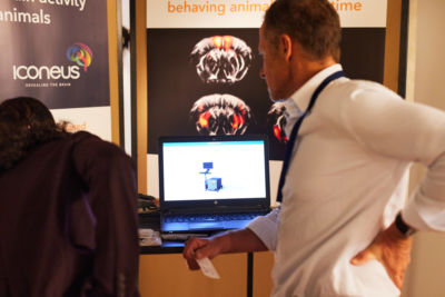
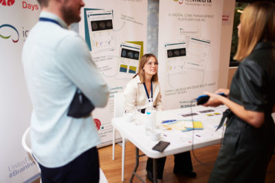
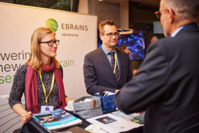
Innovation Hall - Showcase & Posters
The Brain Innovation Hall was the place to be to showcase and discuss your innovation while networking with the various stakeholders in the heart of Brussels.
- The Innovation Hall held:
- Poster Presentations – innovators in the research/ideation phase
- Innovation Showcase - tangible innovations (e.g. device, technology, software) to showcase
Innovation Showcase
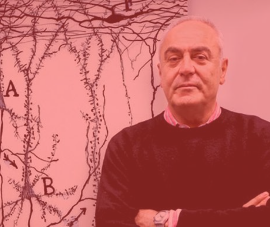
Building Human Induced Nigrostriatal Microcircuits to Study Parkinson’s Disease (Brain On Chip)
Modern medicine is pushing the boundaries of human lifespan and extending the service life of our organs. An exception to this is the brain. The obsolescence of the brain comes in many flavors, and mainly affects two different functions: movement and cognition. Parkinson’s disease (PD) and related disorders impair both. <!–more–> These neurodegenerative diseases remain elusive to the relentless progress in medicine. Some symptoms of PD can be mitigated but we still cannot prevent nor modify the progression of the disease. The main reason why we have not found a cure to PD yet is that we cannot study patient’s brains during the initial stages of the disease. To overcome this limitation, we are collaborating with Imec to generate patient-specific mini-brains on top of CMOS-based multielectrode arrays. Our aim is for Parkinson’s to be objectively defined by solid molecular criteria, rather than by the typical movement symptoms that are currently used to diagnose the disease. This will eventually enable researchers to develop targeted medical interventions that effectively slow down or even stop the disease based on molecular and genetic pathways. The new tools will allow the stratification of patients and the testing of new, personalized therapeutic approaches. We can also apply them to other neurodegenerative diseases, including Amyotrophic Lateral Sclerosis (ALS) and Alzheimer’s.
Carles Calatayud Aristoy, VIB-KU Leuven, Leuven, Belgium
ADMIRE: Alzheimer's Diagnosis via innovative Multimodal Imaging technology of the Retina
Although Alzheimer’s disease (AD) is the number one dementia cause, the currently available diagnostic techniques do not allow preclinical diagnosis or screening due to their high cost, limited resolution and invasiveness, resulting in delayed treatment onset and unsuccessful drug development.
Non-invasive high-resolution retinal imaging tools offers a unique opportunity to overcome the present limitations. ADMIRE is developing a retinal imaging set-up (hardware, image acquisition protocols, and analysis algorithms) for researchers and clinicians using a multimodal approach in which several potential biomarkers are combined: aggregated amyloid beta (Aβ) deposition (using hyperspectral specific filters on microchips integrated into a user-friendly compact camera), neuro-retinal atrophy and (micro)vascular changes (using OCT and fundus images). Prof. Stalmans´ team, in collaboration with IMEC and VIB, was among the first centers worldwide to perform clinical HSRI for AD research with her research leading to evidence for the potential of in vivo HSRI as a biomarker of brain Aβ. Impact on the brain community: The strategic clinical research that is conducted within ADMIRE has an enormous opportunity in terms of translational potential application and utilization objectives for the brain community. ADMIRE will provide a multimodal detection platform for i) population-level preventive and clinical screening, ii) patients identification for clinical trials of drug treatments, iii) patient stratification, and iv) patient monitoring of disease progression, supporting the standard clinical management of dementia.
Dr Sophie Lemmens, Research Group Ophthalmology, KU Leuven, Leuven, Belgium
PhD Jenny Ceccarini, Mission Lucidity, Leuven, Belgium; KU Leuven Brain Institute, Leuven, Belgium
DeepEn: Hair-thin Endoscopes for Deep Brain Imaging
Over the past decade, in-vivo microscopy has enabled many groundbreaking discoveries in different fields of neuroscience, such as behaviour, cognition, and perception. In-vivo microscopy helps us to better understand disorders such as Alzheimer’s, Parkinsons‘, stroke and epilepsy. However, state-of-the-art devices that neuroscientists use for deep-brain microscopy can cause significant damage to the living brain tissue because of their physical dimensions. This tissue damage distorts the brain functions under study. DeepEn is a start-up that develops hair-thin endoscopes for deep-brain microscopy. With this device, neuroscientists can observe processes anywhere in the living brain with sub-micron resolution. The hair-thin probe consists of a single optical fibre, which reduces tissue damage by more than 10 times, compared to existing solutions. The single-fibre endoscope will also make it much easier for researchers to successfully address the brain region of interest. This reduces the risk of wrongly positioning the probe, which saves time and reduces the quantity of animal models required for an in-vivo study. In neuroscientist’s experimental setups, the hair-thin endoscope is a unique and powerful tool to investigate neural connectivity, plasticity, and the neural circuitry. DeepEn’s mission is to support researchers and medical practitioners in discovering, developing, and applying the tools for diagnostics and treatment of brain disorders. DeepEn’s goal is to establish its endoscopes as the primary solution for deep brain imaging. The long-term vision of the start-up is to advance the minimally invasive technology towards applications in medicine.
Website: www.deepen.tech
Hana Cizmarova, Application Management, Leibniz Institute for Photonic Technology, Jena, Germany
Epicranial electrical stimulation for memory enhancement
Many people suffer from cognitive decline caused by hippocampal dysfunction due to neurodegeneration, yet there is no effective medical intervention available. Nonetheless, recent studies have shown a potential role for neuromodulation for slowing down cognitive decline. Existing neuromodulation techniques such as DBS or transcranial alternating current stimulation have several limitations that limit its wide-spread applicability.
We propose a novel minimally invasive method to stimulate deep brain regions involved in memory by means of epicranial electrical stimulation (ECS).
Our preclinical behavioural and functional imaging studies already showed a significant memory improvement and selective hippocampal activation, demonstrating that 40 Hz ECS may be an effective minimally-invasive approach to improve memory. In parallel, we are developing a fully implantable electrical stimulator connected to electrodes implanted on the skull of the patient, with the flexibility in stimulation patterns necessary to reach deeper brain structures involved in memory. This device will be controlled by means of a transcutaneous data link. Since our approach is significantly less invasive than for example DBS, it is in our opinion the only approach viable for treatment so early in the disease. Impact on the brain community: The potential impact of a small implantable device capable of slowing down cognitive decline is massive, including not only patients with mild cognitive impairment but also will be usable in a large number of patients with neuropsychiatric disorders where pharmacological treatments fail and DBS is considered to be too invasive (e.g. depression, chronic pain, Parkinson’s).
Dr. Hannes Heylen, Interuniversity Micro-Electronics Center (IMEC), Leuven, Belgium; Psychiatry, KU Leuven; University Hospitals Leuven; University Hospitals Leuvenn, Leuven, Belgium; Mission Lucidity, Leuven, Belgium
Prof. Dr. Peter Janssen, Laboratory for Neuro- and Psychophysiology, KU Leuven, Leuven, Belgium; KU Leuven Brain Institute, Leuven, Belgium; Mission Lucidity, Leuven, Belgium
A Multimodal Communication Device to Promote the Mental Health of Cobot Operators with Autism Spectrum Disorder
People diagnosed with autism spectrum disorders (ASD) have low employment rates, even though it is known that they can adapt to a variety of jobs if correct measures are taken. In the context of Industry 5.0, the adoption of collaborative robots (cobot) enables the design of inclusive workplaces for people with ASD. However, the uncertainties and sudden actions may create anxiety on these users, therefore a device that could send out information about the status of the cobot and the production is necessary. The present work introduces a modular, complementary device designed according to Design-for-All principles, to improve and make easier the interaction between cobot and people diagnosed with ASD in production lines. The information on the status of the cobot and the production is transmitted through visual and auditory signals, in a rapid and intuitive modality. The data from the cobot is transferred continuously via Bluetooth and converted into light signals displayed by parallel LED strips – thus, through an LED Matrix – on the portable ring component which is directly attached to the cobot; and into sound output communicated via a loudspeaker on the stationary base module, which also serves as the charging port. The product is customizable to communicate additional scenarios if demanded, and its flexible conformation makes it adaptable to diverse types of cobot, making even pre-existing work environments user-friendly. The impact of our work is an implemented human-robot interaction promoting mental health, facilitating positive job experiences, and making the workplace more human-friendly and accessible for all.
Carla Dei, Designer, Scientific Institute IRCCS E. Medea, Bosisio Parini, Italy
Matteo Meregalli Falerni
Novel Interactive BRAINTEASER Tools for Amyotrophic Lateral Sclerosis (ALS) and Multiple Sclerosis (MS) Management
The BRAINTEASER Project (BRinging Artificial INTElligence home for a better care of Amyotrophic lateral Sclerosis and multiple sclERosis) integrates detailed clinical datasets with novel personal health, activity, lifestyle, habitual/behavioural, and environmental data collected using commonly available sensing/IoT devices and the demonstrated first release of developed interactive tools for disease monitoring and management.In this initial release, the mobile app. for patients and informal caregivers and the web tool for clinicians support the key required functionalities of: 1) quantitative and qualitative information collection, consolidation and fusion (of heterogeneous data from IoT wearable trackers and environmental sensing networks, combined with data from digitalized standardized and innovative evolving instruments and questionnaires for ALS and MS (and comorbidities) clinical evaluation and remote disease progress assessment), and 2) elementary multimodal disease management, monitoring and assistance in daily patient and caregiver needs in remote and clinical settings, enabling follow-up, symptoms and health issues resolving, and feedback from the clinicians and caregivers to the patient through various interactive GUI types.The demonstrated tools are being devised and developed embracing an agile user-centred multidisciplinary co-design approach, accounting for the specific technical, medical (including psychological/cognitive) and societal needs of the users.Proof-of-concept of their validated use in real clinical settings on 4 study sites (Lisbon, Madrid, Pavia & Turin) aims to provide quantitative evidence of benefits of leveraging AI models in healthcare pathways for dire neurodegenerative diseases, to support the transition of current care approach from reactive to predictive, towards a healthier and more fulfilling life as long as possible.
Vladimir Urošević, Research & Development Manager, Belit Ltd. Belgrade, Belgrade, Serbia.
FOXIE Eye Tracking system
Purple Gaze helps neurologists rapidly assess and monitor brain disorders in an affordable and non-invasive way using eye movements. Signature abnormalities in eye movements are neural biomarkers of Alzheimer’s Disease, Multiple Sclerosis and Autism Spectrum Disorder. Most of the underlying science is not new and can be traced back to the mid-’60s, but only recent advances in AI algorithms allowed us to build our FOXIE Eye Tracking system that captures 200.000 high-quality data points in just 5 minutes. FOXIE is a revolutionary AI technology because it reduces prohibitively expensive, room-sized and hard-to-operate medical equipment to a portable USB device that can be plugged into any modern computer and used by non-professionals without extensive training. This opens up a new frontier for collecting high-quality eye movements data at scale that will be used as neural biomarkers and drastically improve our understanding of both healthy and pathological brain functioning.
Kirill Korotaev, Co-Founder & CEO, Purple Gaze
Purple Gaze | Utrecht, Netherlands
BrainTrip Dementia Index (BDI) – an AI-optimized EEG-based biomarker that can detect dementia in its early stages and be used to measure its progression
There are currently no scalable or affordable methods for diagnosing dementia at its pre-symptomatic stage, which is generally the only phase when pharmaceutical treatment and behavioral therapies can help postpone mental decline. 50% of all dementia cases never get diagnosed because current methods are expensive, invasive, and only a few can diagnose dementia at an early stage. Additionally, the inaccuracy of current methods makes it virtually impossible to monitor the progression of the disease. BrainTrip has developed a novel AI-optimized EEG-based biomarker for dementia called the BrainTrip Dementia Index (BDI), which can discern early symptoms of the disease at a quick and scalable pace. BrainTrip’s algorithm can detect subtle EEG changes early on, having been developed on high-quality automatically processed EEG data specifically to detect early-stage dementia. BrainTrip’s solution for detecting early-stage dementia is based on a 15-minute EEG test with an accuracy of 90% and can be used by minimally trained staff at any facility. General practitioners can use the BDI as a quick and cheap screening test to optimize referrals. The main benefit of the BDI for neurologists is the ability to monitor disease progression and optimize treatment for each patient. The BDI can help pharmaceutical developers of anti-dementia drugs reduce clinical-trial costs and duration and determine the effectiveness of newly developed drugs. Patients who receive a timely diagnosis can start taking medication earlier (possibly 2 years in advance), which arguably reduces the incidence of injuries and postpones disease progression.
Jurij Dreo, CTO, BrainTrip
BrainTrip | Naxxar, Malta
EEG-based test for cognitive decline screening
Ageing of the European population represents a heavy burden for society, the care and health system in addition to patients and their families as it can be associated with pathological cognitive decline. In some cases, cognitive decline can be slowed down if treated timely. Today, there is no simple and cost-effective solution in primary care for assessing cognitive status.
The objective of our innovation is to provide physicians with the means to make data-driven informed clinical decision in order to make the right patients benefit from interventions that can slow down cognitive decline.
Based on our previous work on neurodegenerative diseases we developed an electroencephalography (EEG)-based brain test that is easy-to-use and cost effective, providing objective physiological markers for cognitive profiling that can help primary care professionals to quickly assess cognitive impairment at an early stage. Impact on brain community as a whole: Our solution has the potential to be easily deployed in primary care. It will impact clinicians, allowing them to detect signs of cognitive decline earlier and faster, in turn affecting the patients’ and their families’ quality of life by providing preventive interventions that will help them remain healthy longer. This will benefit society by extending the amount of year a person remains active and reducing expenses of public health systems by delaying onset of gravest stages of mental diseases. Science and industry will also be impacted as impact of new treatments on cognition could be readily assessed using our system.
Dr Claire Braboszcz, Starlab SL
Clinical-level service for predicting cerebral blood flow
NEAR Brain Inc. is a startup that provides artificial intelligence solutions in the healthcare field. Our mission is to create attractive prediction services by AI-driven digital twin platforms. The product we want to implement is 3D cerebral blood flow simulation models that predict the speed and the pressure of brain blood flow. Recently, new methods for obtaining high-resolution images related to brain imaging have been introduced. However, such imaging techniques have been limited in generating 3D images. In case of brain vessel related diseases like aneurysm, the blood flow speed and pressure are important as well as its geometry. However, the current technology only can provide 3D geometry. To innovate the limited application of MRI images, we plan to provide 3D blood flow simulation model. It makes it possible to predict blood flow characteristics of patients. Furthermore, the risk of neurosurgery can be quantitatively measured and evaluated by the data.
Taerin Lee, CEO, NEAR Brain
NEAR Brain Inc. | Seoul, Republic of Korea
MetaCell Cloud Pharma: integrate proprietary dataset, public multi-omic ontologies, and realistic computational models to advance drug discovery intelligence.
Drug discovery for neurological diseases is an incredibly challenging and expensive business, with ~$102B USD spent in 2021 across all drug discovery initiatives. The optimisation of drug discovery processes is a major unresolved challenge of pharmacological companies. Drug Discovery can be accelerated via the integration of multi-omic data from different ontologies with realistic and explainable AI-models.
We are building a cloud-based solution that allows scientists to aggregate publicly available multi-omic data coming from different curated ontologies with their own proprietary data. Through ingestion this knowledge will then be semantically augmented and also linked to a neuronal phenotype by realistic neuronal network simulations to accelerate drug discovery.
MetaCell Cloud Pharma increases the visibility, searchability and accessibility of information across pharmas to enhance collaboration but also to simplify building applications that will pull information together from multiple sources. By offering a centralized interface for processed data encompassing several projects and related datasets MetaCell Cloud Pharma will allow scientists to find and visualize genomic and phenotypic data of interest and to use cutting edge realistic computational models to discover quantitative links between individual neuronal biophysical properties and neurological disorders. Impact on the brain community: MetaCell Cloud Pharma will guide scientists in the discovery process of new molecules to treat neurological disorders.
Paolo Bazzigaluppi, Principal Scientist, MetaCell, Cagliari, Italy
Towards Treatment of Psychiatric and Neurological Diseases by Targeting Gamma Deficits
Drug discovery for neurological diseases is an incredibly challenging and expensive business, with ~$102B USD spent in 2021 across all drug discovery initiatives. The optimisation of drug discovery processes is a major unresolved challenge of pharmacological companies. Drug Discovery can be accelerated via the integration of multi-omic data from different ontologies with realistic and explainable AI-models.
We are building a cloud-based solution that allows scientists to aggregate publicly available multi-omic data coming from different curated ontologies with their own proprietary data. Through ingestion this knowledge will then be semantically augmented and also linked to a neuronal phenotype by realistic neuronal network simulations to accelerate drug discovery.
MetaCell Cloud Pharma increases the visibility, searchability and accessibility of information across pharmas to enhance collaboration but also to simplify building applications that will pull information together from multiple sources. By offering a centralized interface for processed data encompassing several projects and related datasets MetaCell Cloud Pharma will allow scientists to find and visualize genomic and phenotypic data of interest and to use cutting edge realistic computational models to discover quantitative links between individual neuronal biophysical properties and neurological disorders.
MetaCell Cloud Pharma will guide scientists in the discovery process of new molecules to treat neurological disorders.
Marco Taranta, Business Developer and Mai Nguyen, CEO, OptoCeutics ApS
OptoCeutics ApS | Copenhagen, Denmark
Tackling debilitating CNS diseases using DREADD: Development of a modified receptor that allows pharmaceutical targeting of specific neuronal populations
Vascular Research Group (VRG) has started the development of a new class of therapeutic tools to treat debilitating CNS disorders via inserting engineered “designer” receptors into specific neurons in the brain. The aim of this project is to create a safe, precise and effective alternative for deep brain stimulation (DBS), consisting of a single surgery without any remaining devices followed by chronic pharmacological treatment.
Our project utilizes the so-called DREADD (Designer Receptors Exclusively Activated by Designer Drugs) technology. DREADD receptors can only be activated via their specific, therapeutically applied “designer drugs”. On one hand, the inactivity of these receptors without the “designer drug” offers a safe and individually adjustable treatment. On the other hand, specific activation of these receptors would allow a precise pharmacological modulation of malfunctioning neurons or neuronal circuits without affecting other neuronal processes in the brain – a highly desired goal that is practically impossible to reach with traditional pharmacotherapies.
Our planned delivery method is local microinjection of DREADD-containing vectors (AAV or other), mimicking both the surgical method and mechanism of action of DBS. It is a widely used and extraordinarily effective method to treat various debilitating CNS diseases. However, in contrast to DREADD technology, DBS requires chronic implantation of an electrode and is unable to distinguish between neurochemically different neuron populations. In contrast, using DREADD technology it is feasible to target a neurochemically defined neuron population, offering greater specificity, hence better safety/efficacy profile.
Tamás Kitka PhD
BD and Project Manager, Vascular Research Group
Website: https://vrglab.com
Poster Presentations
Glial cells as potential targets for novel anti-epileptic drugs
Epilepsy is a chronic neurological disorder leading to recurrent epileptic seizures due to abnormal excessive or synchronous neuronal excitation in the brain. More than 70 million people worldwide suffer from epilepsy. Treatment with antiepileptic drugs (AEDs) is the main therapeutic approach. Neuronal structures are primary targets of available AEDs, but about 30% of patients with epilepsy are refractory to AEDs. In recent years, there is a strong evidence confirming the involvement of glial cells including astrocytes and microglia in the pathophysiology of epilepsy.
Understanding AED effects on glia and glia-mediated inflammation may help to explore astrocytes and microglia as potential targets for alternative anti-epileptogenic therapies. For this purpose, in a first step, we aimed to investigate the effects of different AEDs such as tiagabine and zonisamide in an astrocyte-microglia co-culture model of inflammation. Solution & link with innovation: Primary rat astrocytes co-cultures containing 5-10% (M5, “physiological” conditions) or 30-40% (M30, “pathological inflammatory” conditions) of microglia were treated with different concentrations of tiagabine or zonisamide for 24 h. Both drugs differentially regulated the glial properties in the astrocyte-microglia co-culture model of inflammation with regard to glial cell viability, cell-cell communication and microglial morphology. In the next step, the co-culture model can be used for differentiation of new anti-epileptogenic targets.
Development of novel anti-epileptic drugs is challenging. Glial cells with regard to astrocytes and microglia are potential players in future treatment strategies for epilepsy.
Fatme Seval Ismail, Department of Neurology, Klinikum Vest, Recklinghausen, Germany
NIMOCHIP Microfluidic lab-on-a-chip for novel diagnostIcs of neurodegenerative diseases
Amyotrophic lateral sclerosis (ALS) is a severe and fatal neurodegenerative disease characterized by an asymptomatic phase of undetermined duration while it takes on average about nine to 12 months for someone to be diagnosed with ALS. Without a precise and more specific detection, there is no chance for an early and personalized treatment. Early, precise and more specific diagnosis is needed for adequate and personalized treatment. Based on decades of our pre-clinical research with ALS patient sera led us to develop a portable stand-alone clinical “lab on a chip”, NIMOCHIP for disruptive in vitro diagnostics of ALS that could be further expanded to diagnose other neurological disorders with an inflammatory component (e.g. multiple sclerosis, Parkinson’s disease, Alzheimer’s disease, Huntington’s disease,). NIMOCHIP contains multiple chambers with seeded living cells (of animal or human – cell line origin) labeled with fluorescent nanoprobes, that are treated with nanoliter volumes of patient’s samples (purified immunoglobulins G – IgGs from patient sera). NIMOCHIP is using “optics on a chip” technology that could measure a set of physiological fluorescence molecular biomarker signatures (e.g., intracellular calcium, reactive oxygen species – ROS, pH, ion channel responses). The acquired optical signals are analyzed using specialized software to obtain a personalized biomarker signature and to generate a simple diagnostic report for the clinician (the measurements could be performed in a few hours and final results could be given the same day). NIMOCHIP is at TRL4 and a pilot system with flow control is constructed.
Aleksa Marković, Software Developer, Snap One LLC, Belgrade, Serbia
Additional Information
- The presenting author/exhibitor is expected to register and attend the Brain Innovation Days in person and present his/her work.
- Only one abstract can be submitted per presenter.
Please note that for the innovation showcase an additional fee of €100 will be added to the registration fee of the Exhibitor. A high table will be provided in the innovation hall with space for a roll-up or backdrop. Please note that nothing is allowed to be taped on the walls as the building is a protected historical site.
- Abstracts must be submitted online via Oxford abstracts
- All abstracts must be submitted and presented in clear English with accurate grammar and spelling.
- After you start submitting your abstract, you can re-enter the submission link at any time to view and edit your abstract until you submit. Once submitted, you will receive a confirmation email containing the title of your abstract. If you do not receive this confirmation e-mail or have any other questions related to the abstract submission, please contact the team at info@braininnovationdays.eu.
- Abstract submission is open and will close on 31 August 2022.
- Abstract acceptance notifications will be sent in September 2022.
Poster Presentations:
Each poster presenter will be provided with an 1,000 mm (width) x 2,000 mm (height) poster board (poster dimension according to A0-format) and an ample supply of push-pins. The board will be labelled with the preassigned post-it with the title of the poster.
Please note that printing the poster is your responsibility and that all posters should be an A0 format and have a portrait layout in order to fit the poster board (vertical positioning).
Please take into consideration that any charts, diagrams, etc should be enlarged and easily readable on the poster.
Innovation Showcases:
- Please note that you will be provided a high table and chair. If you need any additional materials (e.g. screen, extra chairs, etc.) you may bring these yourself or order at an additional cost through the venue – please contact the Brain Innovation Days team to let us know your needs as soon as possible: info@braininnovationdays.eu
- Tables will be preassigned for each innovation showcase by the title of the submission.
We encourage all successful applicants to register soon.